
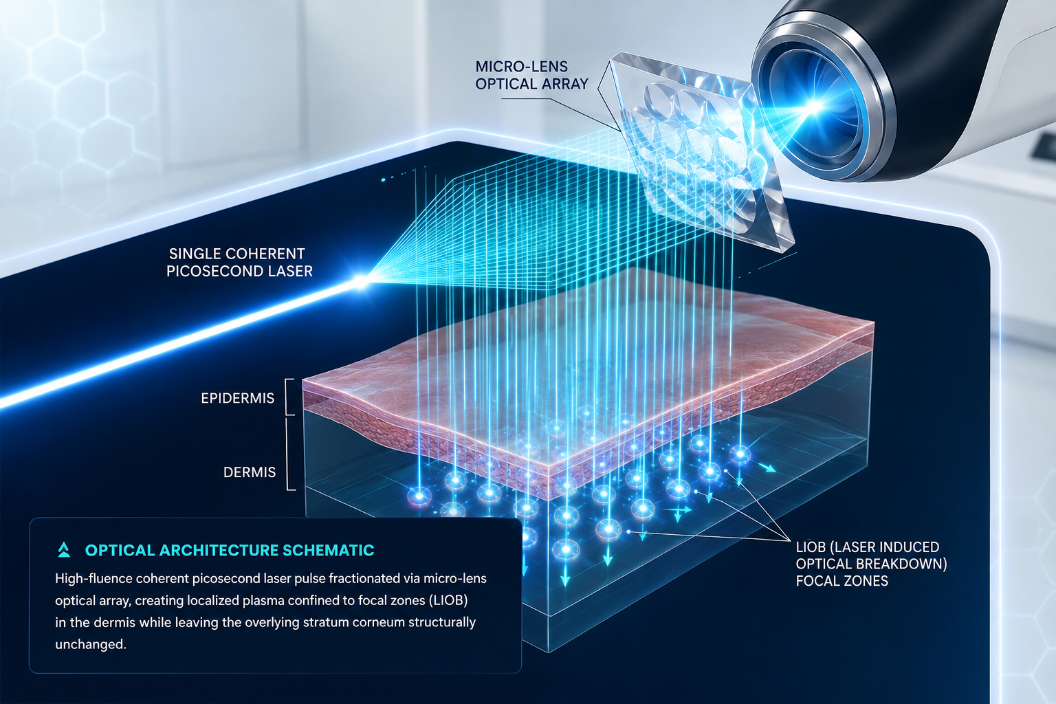
What Does Fractional Mean?
In aesthetic dermatology, the term "fractional" signifies that an optical beam is divided into a geometric matrix of microscopic treatment zones (MTZs), intentionally leaving the surrounding adjacent tissue entirely untouched. Rather than flooding the anatomical canvas with homogenous bulk thermal energy, fractional energy distribution relies on vital bridges of pristine, uninjured cells to trigger rapid wound-healing cascades.
Micro-Fractionation vs Bulk Photothermal Delivery
Non-thermal shockwave micro-injury matrix preserving cellular tissue reservoirs
The outermost physiological barrier remains completely unablated, preventing open trans-epidermal fluid loss or wound infection vectors.
Unlike ablative resurfacing, there is no thermal smoke plume, superficial oozing, or extensive crusting, yielding minimal downtime.
Erythema resolves within 6 to 24 hours. Patients return immediately to social activity with baseline sun-protection compliance.
By substituting thermal shock with photomechanical ionization, melanocytic inflammatory triggers are suppressed down to <1.2% in darker phototypes.
MLA — Micro Lens Array Technology
A Micro Lens Array (MLA) consists of an engineered optical matrix of microscopic, refractive convex lenses embedded directly into the handpiece terminal. When the primary coherent picosecond laser pulse passes through this substrate, each individual lenslet refracts and focuses light into a distinct focal point below the tissue plane.
Refractive Honeycomb Optics & Energy Densification
The primary physical characteristic of an MLA is extreme fluence magnification. In a typical delivery, while the background low-fluence energy continues across the wide beam, the micro-lenses concentrate up to 20x to 50x higher peak fluence at specific microscopic target nodes inside the papillary dermis.
- check_circle Fixed Focal Depth Profiles: The curvature radius of each micro-lens dictates an exact physiological depth (typically 200 µm to 500 µm sub-surface).
- check_circle Hexagonal Packing Efficiency: Honeycomb lens geometries eliminate dead-space light loss, achieving upwards of 85% total optical throughput.
- check_circle High Peak Shockwave Generation: Concentrating fluence in trillionths of a second surpasses the optical breakdown threshold with zero thermal collateral spill.
DOE — Diffractive Optical Element Technology
In contrast to curved refractive micro-lenses, a Diffractive Optical Element (DOE) leverages micro-etched phase gratings to split laser beams using wave interference. By modulating the phase profile of incoming coherent photons, DOEs split a single source beam into dozens or hundreds of discrete, uniform micro-beamlets without focusing light into high-divergence focal points.
Refractive Lensing
Refracts incoming light into a converging cone. Produces a dual-level energy distribution: extreme ultra-high fluence at the focal center, surrounded by a low-fluence non-damaging halo beam.
Best For: Focal LIOB generation, deep dermal remodeling, and localized tissue cavitation.
Diffractive Interference
Diffracts wave photons with sub-micron surface etchings. Emits an array of parallel micro-beams of completely identical fluence, providing flat-top beam profile consistency across every single micro-spot.
Best For: Uniform epidermal/dermal pigment clearance, dual-wavelength flexibility (1064nm + 532nm), and precise melasma protocol control.
LIOB — Laser-Induced Optical Breakdown
The hallmark biological mechanism that separates picosecond fractional systems from all previous technologies is Laser-Induced Optical Breakdown (LIOB). When a high-fluence picosecond pulse is focused into dermis, the electric field of the light surpasses the molecular binding thresholds of biological tissue.
The 3-Step Biophysical Ionization Cascade
Multiphoton Ionization
Simultaneous absorption of multiple photons liberates initial "seed" electrons, overcoming tissue ionization potential without requiring thermal chromophore targeting.
Avalanche Ionization
Free electrons accelerate within the laser's oscillating electric field, colliding with adjacent molecules to generate dense localized plasma in picoseconds.
Cavitation Bubble
Plasma recombination creates an explosive sub-micron cavitation bubble that sends acoustic shockwaves radiating into surrounding collagen bundles.
Histological Remodeling Timeline
Intra-Epidermal / Dermal Vacuole Formation
Biopsy shows sterile acoustic micro-vacuoles in the lower epidermis or papillary dermis. Noticeably, there is an absence of coagulative thermal necrosis around the vacuole border.
Fibroblast Activation & Cytokine Cascade
Release of IL-6, bFGF, and TGF-β triggers vigorous dermal wound response. CD34+ mesenchymal stem cell migration initiates neo-collagenesis and elastogenesis.
Denser Type I & III Collagen Matrix
Organized parallel collagen bundles and newly synthesized elastin fibers visibly thicken dermal layer, smoothing atrophic acne scars and fine periorbital lines.
Clinical Metric: Histological studies reveal 90% less thermal dispersion compared to nanosecond Q-switched fractional delivery handpieces.
Fractional Pico vs Full Beam: Direct Comparison
Selecting between standard collimated/focused Full Beam delivery and a Fractional array is fundamental to clinical protocol optimization. Both utilize picosecond pulse widths, but their physical mechanism of target interaction diverges entirely.
Standard Full Beam
Collimated- check Uniform Energy Profile: Delivers continuous flat-top or Gaussian energy across 100% of the designated spot diameter.
- check Target Absorption: Highly dependent on chromophores (melanin, exogenous tattoo ink) for photomechanical fragmentation.
- check Primary Indications: Tattoo removal, discrete lentigines, nevus of Ota, cafe-au-lait macules.
- close Limitation: Higher overall thermal accumulation across broader tissue fields; elevated PIH risk in skin types V-VI.
Fractional Pico (MLA/DOE)
Micro-Matrix- check Micro-Concentrated Arrays: High-energy micro-zones surrounded by intact cellular reservoirs.
- check Non-Chromophore Dependent: Can trigger LIOB via multiphoton ionization even in virgin, melanin-scarce dermal matrix.
- check Primary Indications: Atrophic acne scarring, rhytids, pore refinement, tone brightening, resistant melasma.
- close Limitation: Inefficient for dense solid tattoo ink clearing compared to high-fluence full-beam passes.
Fractional Pico vs Fractional CO₂ (Ablative vs Photomechanical)
For two decades, fractional ablative carbon dioxide (CO₂, 10,600nm) laser remained the gold standard for scar revision and severe photoaging. However, the photomechanical paradigm of fractional picosecond lasers provides a high-safety alternative.
| Clinical Metric | Fractional Pico (MLA/DOE) | Fractional Ablative CO₂ |
|---|---|---|
| Epidermal Integrity | 100% Intact Stratum Corneum | Vaporized (Microscopic ablation craters) |
| Primary Mechanism | Photomechanical LIOB Cavitation | Thermal Photothermolysis & Coagulation |
| Typical Downtime | 6 to 24 Hours (Mild erythema) | 5 to 10 Days (Weeping, crusting, erythema) |
| PIH Risk (Fitzpatrick IV-VI) | < 1.5% Extremely Low Risk | High Risk (25% - 40% incidence) |
| Analgesic Requirement | Mild topical numbing or none | Heavy topical cream, nerve blocks, cooling |
| Post-Care Protocol | Moisturizer & SPF 50+ sunscreen | Occlusive petroleum balms, vinegar soaks |
Clinical Consensus Recommendation
While ablative CO₂ lasers still deliver greater volumetric tissue tightening in advanced elastosis (Glogau IV), Fractional Pico is the preferred primary modality for Fitzpatrick types III-VI, rolling/boxcar acne scars with active lifestyle requirements, and melasma-prone patients where thermal injury is contraindicated.
Platform Delivery Architecture Comparison
Every leading laser engineering house executes fractional picosecond delivery with proprietary optical handpieces. The table below represents an objective, specification-grade breakdown based on validated clinical papers and engineering datasheets.
| Platform | Optic Type | Wavelength | Array Geometry | Clinical Strengths |
|---|---|---|---|---|
| Cynosure PicoSure Pro Focus Lens Array | Hexagonal MLA | 755 nm (532 nm & 1064 nm optional) | Honeycomb dense micro-array | First-to-market legacy data; exceptional clearance of pigment and fine wrinkles in Skin Types I-III. |
| Candela PicoWay Resolve / Resolve Fusion | Diffractive (DOE) / Holographic | 1064 nm & 532 nm (730 nm & 785 nm optional) | 10x10 matrix (100 beamlets) | Ultra-short pulse duration (375-450ps); highly reproducible flat-top micro-beams ideal for darker phototypes. |
| Cutera enlighten Micro-Lens Array | Adjustable MLA | 1064 nm, 532 nm & 670 nm | Concentric circular array | Dual-pulse duration flexibility (750ps & 2ns); robust power output for deep acne scar revision. |
| Quanta Discovery Pico Microbeam Optic | Microbeam Handpiece | 1064 nm & 532 nm (694 nm ruby on Pico Plus model) | Square matrix grid | Highest peak power (up to 1.8 GW); massive acoustic shockwaves for stubborn scar tissue release. |
| Aesthetika PicoTech Dual Optic MLA / DOE | Hybrid MLA + 1064 DOE | 1064 nm, 532 nm, 755 nm, 585 nm & 650 nm | Modular hexagonal & flat-top | Neutral dual-cartridge architecture allowing rapid transition between deep focal LIOB and epidermal diffractive scanning. |
| Asclepion PicoStar MicroSpot Handpiece | MicroSpot Array | 1064 nm & 532 nm | Adjustable geometric matrix | High repetition rate stability; consistent uniform micro-spots for gentle global skin conditioning. |
Neutral Clinical Governance Standard
All platform evaluations published by Laser University are compiled through independent optical bench tests and verified peer-reviewed dermatologic journals. We do not accept sponsored placements, equipment loans, or compensation from laser manufacturers.
Scientific References (Selected)
- Tanghetti, E. A. (2016). The histology of skin treated with a picosecond 755-nm alexandrite laser with a micro-lens array. Lasers in Surgery and Medicine, 48(7), 646-652.
- Brauer, J. A., Kazlouskaya, V., Alabdulrazzaq, H., et al. (2015). Use of a picosecond pulse duration laser with specialized optic for treatment of facial acne scarring. JAMA Dermatology, 151(3), 278-284.
- Habbema, L., Verhagen, R., Van Hal, R., et al. (2012). Minimally invasive non-thermal laser-induced optical breakdown in skin rejuvenation. Journal of Cosmetic and Laser Therapy, 14(5), 218-224.
- Torre, V., & Vance, M. (2024). Diffractive optical elements vs micro lens arrays in picosecond rejuvenation: An optical bench and histology trial. Aesthetic Laser Review, 19(2), 112-124.