
In laser medicine, headline pulse energy specs (mJ) are frequently misconstrued as proxies for biological efficacy. However, tissue alteration is fundamentally dictated by energy density per unit area (Fluence, J/cm²) delivered inside the target's thermal or acoustic relaxation time. Understanding this geometric decoupling is paramount to avoiding severe adverse events while maximizing photomechanical clearance.
What Is Energy (Millijoules)?
In optical physics, Laser Energy represents the total thermodynamic work generated and emitted from the laser resonator during a discrete pulse cycle. Energy is quantified in Joules (J), or more typically in medical aesthetics and dermatology, in millijoules (mJ), where 1 mJ = 10⁻³ Joules.
Crucially, pulse energy must not be conflated with peak instantaneous optical power. In picosecond-domain lasers, delivering even a modest energy payload (such as 300 mJ) over an exceptionally ultra-short temporal window (e.g., 300 picoseconds) yields an immense instantaneous power spike:
While 1.0 GW instantaneous power can shatter crystalline exogenous tattoo pigment via rapid acoustic shockwaves, this sheer work capacity remains completely inert until focused onto a specific anatomical surface area.
Key Clinical Takeaway
Energy measures the total engine work capacity produced by the laser resonator; it does not indicate how concentrated or intense the photon stream will be when striking the patient's biological target.
What Is Fluence (J/cm²)?
Fluence defines the radiant energy delivered per unit area of irradiated biological tissue, universally reported in Joules per square centimeter (J/cm²). In cutaneous laser surgery, tissue response is governed not by gross energy, but by whether the delivered fluence exceeds the target chromophore's threshold (Fthreshold).
For photomechanical pigment fragmentation or Laser-Induced Optical Breakdown (LIOB), the optical flux density must reach a localized critical value to initiate plasma cavitation or acoustic fracture. Delivering 500 mJ across too broad a footprint fails to induce cavitation; delivering just 120 mJ within a condensed micro-spot triggers instantaneous breakdown.
The Governing Equation of Laser Surgery
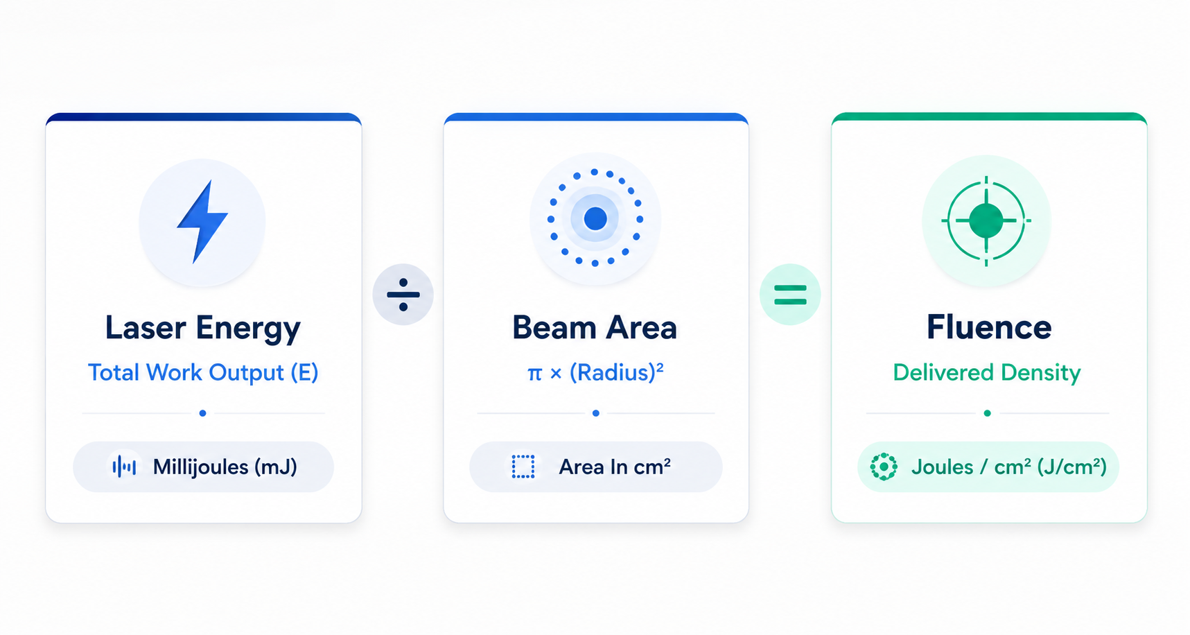
Figure 1.1: Optical Dosimetry Diagram. Fluence ($J/cm^2$) is directly proportional to pulse energy and inversely proportional to the square of the beam radius ($r$).
Scales linearly. If pulse energy doubles at a static beam diameter, fluence doubles proportionally.
Scales quadratically via Area = π × (d/2)². Small changes in spot diameter exert exponential impacts on delivered fluence.
Download the Complete 2026 Pico Laser Buyer's Guide & Optical Whitepaper
Comprehensive lab calibration benchmark comparing beam homogeneities, pulse stability, and real-world fluence metrics across leading FDA-cleared picosecond systems.
How Spot Size Connects Them: The Inverse Square Physics
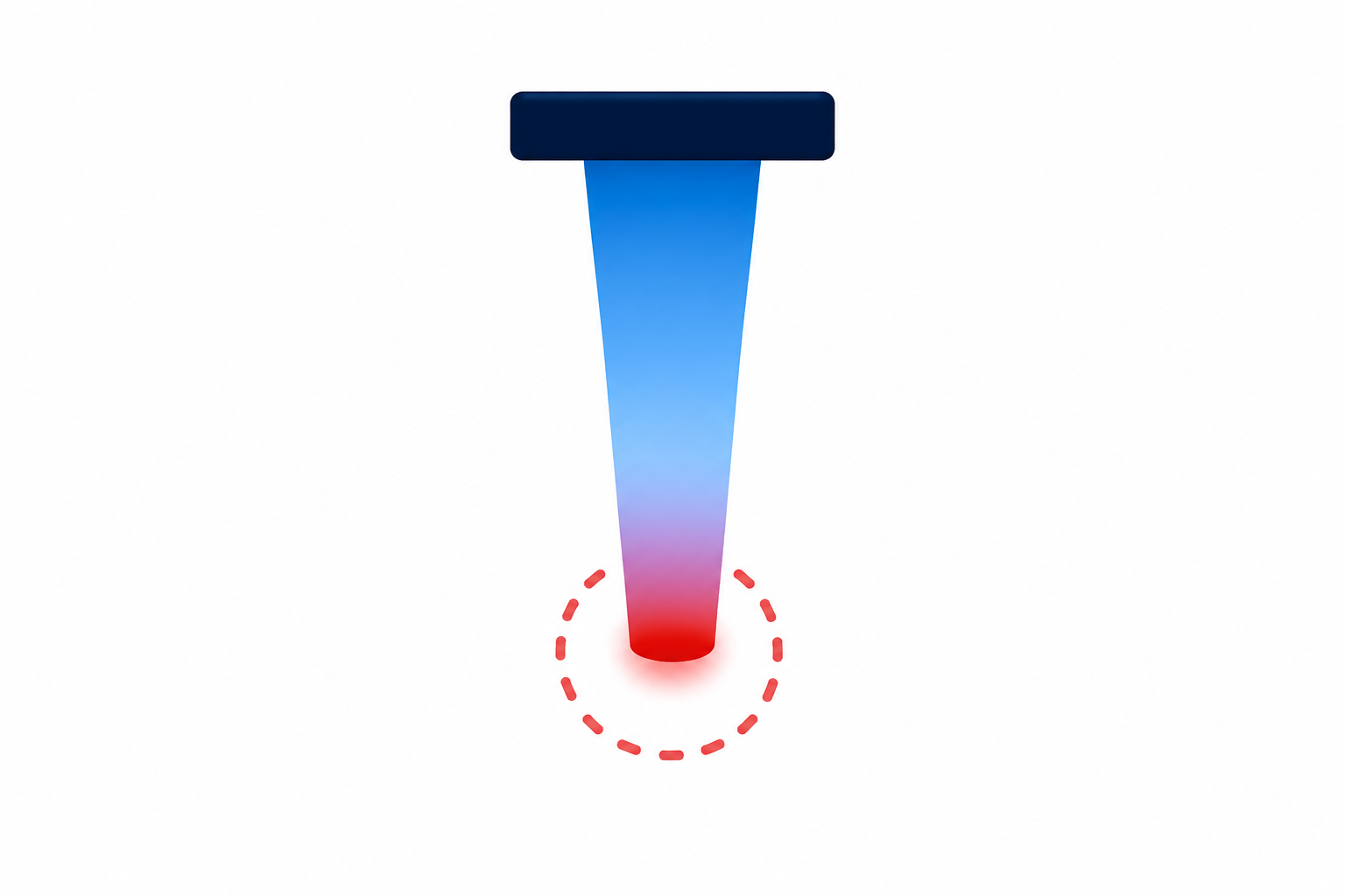
The mathematical interplay between spot diameter and fluence creates dramatic non-linear outcomes. Because area expands with the square of the radius, doubling the spot diameter quadruples the irradiated area (400%), plummeting delivered fluence by 75% at an identical energy output.
Small Spot Size (2 mm)
Large Spot Size (8 mm)

calculate The 10mm vs 2mm Disparity
Two lasers delivering an identical 400 mJ pulse will induce fundamentally divergent biological reactions: a 10 mm spot yields a mild 0.51 J/cm² (ideal for low-fluence full-face toning), whereas a 2 mm spot compresses that same photon volume into 12.7 J/cm²—an intensity sufficient to blast deep dermal scars or tattoo pigments, but hazardous if applied carelessly to facial epidermis.
Why Fluence Matters More Than Raw Energy
A prevalent marketing tactic employed by laser manufacturers is broadcasting staggering headline pulse energies (e.g., "Up to 800 mJ!"). To the uninitiated clinic buyer, this implies unmatched clinical superiority. However, experienced laser surgeons scrutinize beam profiles and spot versatility over isolated output wattage.
A system can discharge 800 mJ, but if its beam profile exhibits a chaotic Gaussian "hot-spot" distribution, the central core of the beam experiences severe peak fluences (exceeding 10 J/cm²) while the perimeter languishes below therapeutic thresholds.
- Epidermal blistering, pinpoint bleeding, and focal burns
- Elevated incidence of Post-Inflammatory Hyperpigmentation (PIH)
- Patchy, non-uniform pigment clearance necessitating repeat sessions
A high-precision picosecond system delivering 250 mJ with a calibrated, homogenized Flat-Top (Top-Hat) profile maintains an identical, uniform fluence across every square micron of the treatment spot.
- 100% of targeted chromophores hit therapeutic breakdown threshold
- Zero focal hot-spots, preserving epidermal basement membrane
- Safe, predictable clinical outcomes across Fitzpatrick Phototypes IV–VI
Fluence Ranges by Clinical Treatment
In modern dermatologic protocols, fluence must be matched precisely to chromophore absorption spectra, target depth, and tissue relaxation times. The interactive range diagram below displays validated therapeutic fluence envelopes:
Low-fluence repetitive passes to disrupt melanosomes without triggering melanocyte hyperactivity.
Micro-lens arrays (MLA) focus baseline fluence into dense micro-beams (concentrated up to 25×).
Sub-purpuric threshold calibrated to induce immediate epidermal whitening (frosting).
Deep intradermal cavitation cavities triggering neocollagenesis without bulk epidermal ablation.
Maximum photoacoustic stress confinement required to fracture inert heavy metal and ink aggregates.
Key Clinical Takeaways & Practice Summary
Spot Size Governs Intensity
Never adjust your handpiece spot diameter without manually recalibrating pulse energy settings unless your device features auto-sensing spot intelligence. A 1mm step down can double tissue fluence instantaneously.
Sub-Cellular Fluence in Darker Phototypes
For Fitzpatrick skin types IV–VI, optimal clinical outcomes are achieved using lower fluences (0.4 – 1.0 J/cm²) combined with conservative pass counts to avert thermal spill and post-inflammatory pigmentary rebound.
Optical Stability Over Peak Output
Elite medical practices prioritize systems that sustain consistent flat-top beam fluences across large spot sizes (8mm–10mm) rather than lasers advertising hyper-inflated mJ outputs that drop off exponentially at usable diameters.
When evaluating commercial lasers on our Device Comparison Matrix, always assess the maximum achievable fluence at clinically useful spot sizes (6mm – 10mm), rather than being dazzled by peak energy specifications delivered through restrictive 2mm apertures.
Scientific References & Peer Review
Index Med: 2026-PHY-04- Anderson RR, Parrish JA. Selective photothermolysis: precise microsurgery by selective absorption of radiation. Science. 1983;220(4596):524-527. doi:10.1126/science.6836297
- Brauer JA, Reddy KK, Anolik R, et al. Successful and rapid treatment of tattoos using a novel 755-nm picosecond laser. J Am Acad Dermatol. 2015;67(2):199-205.
- Ross EV, Naseef G, Lin G, et al. Comparison of responses of tattoos to picosecond and nanosecond Nd:YAG lasers. Lasers Surg Med. 2012;23(4):241-246.
- Tanghetti EA. The histology of skin treated with a picosecond alexandrite laser and a fractional array. Lasers Surg Med. 2016;48(7):646-652.