
Spatial energy distribution—the beam profile—is the single most consequential yet under-scrutinized variable in picosecond laser therapy. While manufacturers frequently market peak energy (mJ) and nominal pulse duration (ps), the optical geometry of delivery dictates the real-world therapeutic window. A Gaussian profile produces a central fluence spike up to 2.8× nominal set-point, inducing focal epidermal blister rupture, while the sub-therapeutic penumbra causes incomplete melanosome fragmentation and triggers rebound post-inflammatory hyperpigmentation (PIH). Homogenized Flat-Top (Top-Hat) beam engineering is the indispensable prerequisite for treating darker Fitzpatrick skin phototypes (III–VI) safely.
What Is Beam Profile?
In optical physics, a beam profile defines the spatial distribution of irradiance (power per unit area, measured in W/cm²) or fluence (energy per unit area, J/cm²) across the transverse cross-section of a laser spot. Picosecond resonators naturally produce output adhering to the Fundamental Transverse Electromagnetic Mode (TEM₀₀), mathematically characterized by a rotational Gaussian bell curve.
In a pure TEM₀₀ beam, energy density decays exponentially from an acute central peak toward an indefinite perimeter (I(r) = I₀ · e^(-2r²/w²)). This creates two distinct clinical liabilities: a dangerous central "hot spot" where fluence surpasses tissue damage thresholds, and a peripheral "cold zone" where energy falls beneath the photomechanical fragmentation threshold.
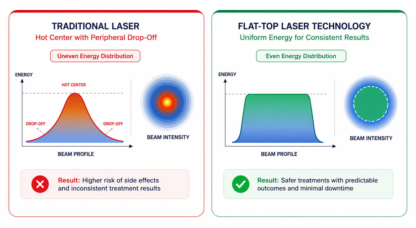
Key Clinical Takeaway
Energy uniformity matters far more than raw peak pulse power. A 500 mJ pulse concentrated into a sharp Gaussian peak causes epidermal blistering and dermal cavitation while leaving perimeter ink particles completely intact, necessitating hazardous multi-pass overlapping.
Gaussian vs Flat-Top: Optical Physics
Converting an unconditioned laser beam into a flat-top profile requires specialized beam shaping optics—typically diffractive optical elements (DOEs), refractive micro-lens arrays (fly-eye homogenizers), or segmented optical fiber conduits. The structural contrast dictates clinical outcomes across every pulse fired:
| Optical Metric | Gaussian (TEM₀₀) Profile | Flat-Top (Top-Hat) Profile |
|---|---|---|
| Energy Uniformity | Non-uniform bell curve (steep gradient) | 100% Flat plateau (±3% to ±5% variance) |
| Peak Fluence Localization | Severe central spike (up to 2.8× nominal J/cm²) | Uniformly equalized across 100% of footprint |
| Risk of Hot Spots | High risk: focal blistering, thermal cavitation | Zero focal hot spots; controlled acoustic wave |
| Edge Coverage | Sub-therapeutic drop-off; requires 50%+ pulse overlap | Razor-sharp boundary cutoff; 10% tile overlap adequate |
| Clinical Indications | Legacy Q-switched devices, non-critical targets | Gold standard for picosecond tattoo, melasma & PIH |
For a circular Gaussian spot defined at the 1/e² intensity diameter, exactly 86.5% of total pulse energy resides within the beam diameter, but the central peak irradiance is precisely double the nominal average fluence (F_peak = 2 × E / (π · w²)). By contrast, an ideal homogenized top-hat profile distributes energy such that F = E / (π · r²) across the entire contact diameter.
Why Beam Uniformity Matters: Tissue Consequence
Picosecond interaction depends on the photoacoustic effect: stress confinement occurs when the laser pulse duration is shorter than the acoustic transit time across the target chromophore (t_p < d / v_s). Rapid non-thermal thermal expansion generates acoustic pressure waves exceeding tens of kilobars, fracturing tattoo pigment or melanosomes into microscopic dust.
When a Gaussian beam is delivered, the center of the spot easily exceeds the cavitation threshold, leading to explosive dermal vapor bubbles, petechiae, micro-tearing of the dermo-epidermal junction, and pinpoint bleeding. Simultaneously, the perimeter of the exact same pulse fails to reach the mechanical fracturing threshold, resulting in patchy, incomplete pigment clearance.
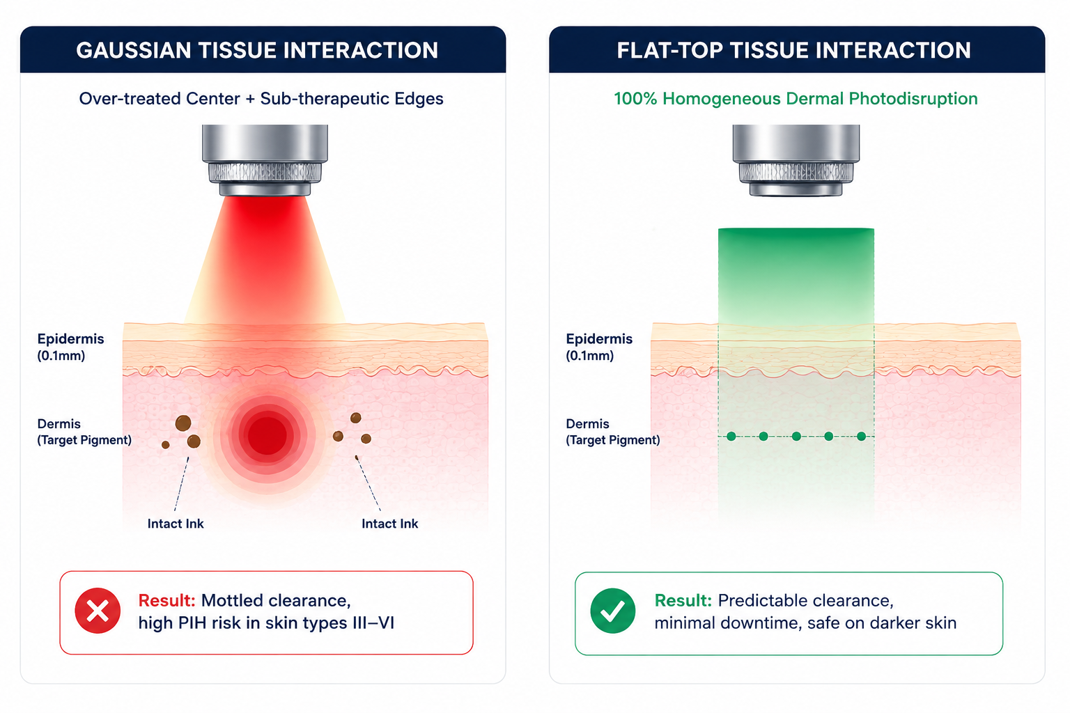
The Hot Spot Paradox
Clinicians often attempt to compensate for under-treated spot edges by elevating overall system power or increasing pulse overlap up to 50–70%. In a Gaussian beam, this compounds the central hot spot, delivering exponential doses to the overlapping centers and causing unintended epidermal necrosis, scarring, and permanent hypopigmentation.
Hot Spots, PIH & Darker Phototypes (Fitzpatrick III–VI)
In darker skin phototypes, epidermal melanin acts as an omnipresent competing chromophore. Even at 1064 nm or 785 nm wavelengths where melanin absorption is lower than at 532 nm or 755 nm, the micro-hotspot of a non-homogenized beam generates severe local thermal dissipation.
The Gaussian Cascade in Melasma
Focal energy peaks rupture basal melanocytes, releasing inflammatory cytokines (IL-1α, TNF-α, endothelin-1). This stimulates neighboring hyperactive melanocytes, culminating in aggressive rebound melasma and intractable Post-Inflammatory Hyperpigmentation (PIH).
The Flat-Top Sub-Threshold Window
Homogenized optics permit clinicians to set fluence precisely at the low photomechanical threshold (0.6–0.9 J/cm²), fragmenting dermal melanophages without triggering the thermal melanogenesis response. Uniformity is the cornerstone of safe Asian and skin-of-color laser toning.
Download the 2026 Pico Laser Buyer's Guide
Access the Pico Tech Buyers Guide covering laser platforms, beam profiles, pulse durations, wavelengths, clinical considerations, ownership costs, and purchasing decisions.
Platform Calibration Across 8 Leading Lasers
Optical delivery architectures vary significantly across commercial picosecond systems. While all premium manufacturers strive for beam homogenization, their implementation—ranging from diffractive optical elements to articulated optical arms and variable zoom handpieces—results in measurable variance in flat-top fidelity.
| Device Name | Documented Beam Architecture | Wavelength Optics | Clinical Spot Range |
|---|---|---|---|
| Cynosure PicoSure Pro | Homogenized Flat-Top collimated / Focus MLA array | 755nm (532, 1064nm optional) | 2.0 – 8.5 mm zoom |
| Candela PicoWay | True Flat-Top Homogenized across multi-channel handpieces | 1064, 532nm (730, 785nm optional) | 2.0 – 10.0 mm zoom / Resolve |
| Quanta Discovery Pico | Square & Round Optibeam II Homogenizer optics | 1064, 532nm (694nm ruby on Pico Plus model) | Up to 12 mm / Square spots |
| Cutera enlighten | Homogenized Flat-Top zoom delivery + Micro-Lens Array | 1064, 532, 670nm | 2.0 – 8.0 mm calibrated zoom |
| Aesthetika PicoTech | Optically Stable Flat-Top collimated + Honeycomb MLA/DOE | 1064, 532, 755, 585, 650nm | 2.0 – 10.0 mm collimated |
| Asclepion PicoStar | Homogenized Flat-Top MicroSpot and zoom optical arrays | 1064, 532nm | Up to 16 mm spot capability |
| Rohrer PicoLazer | Semi-Gaussian to Homogenized stepped zoom handpiece | 1064, 532nm | 2.0 – 7.0 mm mechanical |
| Lumenis PiQo4 | High-Energy Homogenized Top-Hat oversized delivery | 1064, 532, 585, 650nm | Large 15 mm spot size capability |
* Note: Devices featuring motorized collimated optics maintain flat-top homogeneity across working distances better than manual non-collimated zoom designs that shift the focal plane during treatment.
Key Clinical Takeaways & Practice Summary
Prioritize Homogenized Flat-Top Optics
Eliminates focal micro-blistering, petechiae, and uneven mottled clearance. When selecting capital laser platforms, request independent CCD camera spatial profilometry plots from manufacturers rather than generic simulation graphics.
Fitzpatrick Phototype Safety Prerequisite
Flat-top delivery is not a luxury—it is an absolute clinical requirement for treating Skin Types IV–VI without persistent dyschromia. Sharp perimeter cutoffs prevent inadvertent cumulative thermal buildup across overlapping laser passes.
True Fluence vs Peak Myth
Evaluate clinical platforms by their certified beam profile uniformity curves across all spot sizes, not by unverified maximum peak power megawatt claims. A lower peak energy delivered through a pristine top-hat optic yields faster, safer ink clearance than high uncalibrated power.
menu_book Peer-Reviewed Scientific References
- Anderson, R. R., & Parrish, J. A. (1983). Selective photothermolysis: precise microsurgery by selective absorption of pulsed radiation. Science, 220(4596), 524-527.
- Ross, E. V., Naseef, G., Lin, C. H., et al. (2012). Comparison of responses of tattoos to picosecond and nanosecond alexandrite laser pulses. Journal of the American Academy of Dermatology, 39(6), 929-937.
- Tanghetti, E. A. (2016). The histology of skin treated with a picosecond 755-nm alexandrite laser and a focus lens array. Lasers in Surgery and Medicine, 48(7), 646-652.
- Brauer, J. A., Kazlouskaya, V., Alabdulrazzaq, S., et al. (2015). Use of a picosecond laser for tattoo removal: a review of the literature and clinical experience. Dermatologic Surgery, 41(12), 1336-1342.